Tuesday, October 13, 2009

Temporomandibular joint disorder


Temporomandibular joint

Temporomandibular joint disorder (TMJD or TMD), or TMJ syndrome, is an umbrella term covering acute or chronic inflammation of the temporomandibular joint, which connects the mandible to the skull. The disorder and resultant dysfunction can result in significant pain and impairment. Because the disorder transcends the boundaries between several health-care disciplines — in particular, dentistry, neurology, physical therapy, and psychology — there are a variety of treatment approaches.

The temporomandibular joint is susceptible to many of the conditions that affect other joints in the body, including ankylosis, arthritis, trauma, dislocations, developmental anomalies, and neoplasia.


Signs and symptoms

Signs and symptoms of temporomandibular joint disorder vary in their presentation and can be very complex. Often the symptoms will involve more than one of the numerous TMJ components: muscles, nerves, tendons, ligaments, bones, connective tissue, and the teeth. Ear pain associated with the swelling of proximal tissue is a symptom of temporomandibular joint disorder.

Muscles

TMJ diagram

Disorders of the muscles of the temporomandibular joint are the most common complaints by TMD patients. The two major observations concerning the muscles are pain and dysfunction. The dysfunction can present as trismus or limitation of jaw movement ranging from minor to severe. In milder cases, the only representation may be joint sound such as clicking or popping. These symptoms of TMD are often caused by overusage of the muscles of mastication. Common causes include chewing gum continuously, biting habits (fingernails and pencils), grinding habits, and clenching habits.

Most cases of TMJ, however, are not so simple. Deep-space infections with resulting trismus or neoplams about the joint may mimic TMJ dysfunction. Muscle pain can sometimes be associated with trigger points in muscle tissue. These trigger points can be localized by digital palpation, both intraorally and extraorally. This is known as Myofascial pain syndrome.

Any dysfunction of the muscles may cause the teeth to occlude (bite) with each other incorrectly; if teeth are traumatized by this, they may become sensitive, demonstrating one of the many interplays between muscle, joint, and tooth.

Temporomandibular joints

This is arguably the most complex set of joints in the human body. Unlike typical finger or vertebral junctions, each TMJ actually has two joints, which allow it to both rotate and to translate (slide). With use, it is common to see wear of both the bone and cartilage components of it. Clicking is common, as are popping motions and deviations in the movements of the joint. It is considered a TMJ disorder when pain is involved.

In a healthy joint, the surfaces in contact with one another (bone and cartilage) do not have any receptors to transmit the feeling of pain. The pain therefore originates from one of the surrounding soft tissues. When receptors from one of these areas are triggered, the pain causes a reflex to limit the mandible's movement. Furthermore, inflammation of the joints can cause constant pain, even without movement of the jaw.

Due to the proximity of the ear to the temporomandibular joint, TMJ pain can often be confused with ear pain. The pain may be referred in around half of all patients and experienced as otalgia (earache). Conversely, TMD is an important possible cause of secondary otalgia. Treatment of TMD may then significantly reduce symptoms of otalgia and tinnitus, as well as atypical facial pain. Despite some of these findings, some researchers question whether TMD therapy can reduce symptoms in the ear, and there is currently an ongoing debate to settle the controversy.

The dysfunction involved is most often in regards to the relationship between the condyle of the mandible and the disc. The sounds produced by this dysfunction are usually described as a "click" or a "pop" when a single sound is heard and as "crepitation" or "crepitus" when there are multiple, rough sounds

Teeth

Disorders of the teeth can contribute to TMJ dysfunction. Impaired tooth mobility and tooth loss can be caused by destruction of the supporting bone and by heavy forces being placed on teeth. The movement of the teeth affects how they contact one another when the mouth closes, and the overall relationship between the teeth, muscles, and joints can be altered. Pulpitis, inflammation of the dental pulp, is another symptom that may result from excessive surface erosion. Maybe the most important factor is the way the teeth meet together: the equilibration of forces of mastication and therefore the displacements of the condyle.


Precipitating factors

There are many external factors that place undue strain on the TMJ. These include but are not limited to the following:

Over-opening the jaw beyond its range for the individual or unusually aggressive or repetitive sliding of the jaw sideways (laterally) or forward (protrusive). These movements may also be due to parafunctional habits or a malalignment of the jaw or dentition. This may be due to:

  1. Trauma
  2. Repetitive unconscious jaw movements called bruxing.
  3. Malalignment of the occlusal surfaces of the teeth due to dental defect or neglect.
  4. Jaw thrusting (causing unusual speech and chewing habits).
  5. Excessive gum chewing or nail biting.
  6. Size of foods eaten.
  7. Degenerative joint disease, such as osteoarthritis or organic degeneration of the articular surfaces, recurrent fibrous and/or bony ankylosis, developmental abnormality, or pathologic lesions within the TMJ
  8. Myofascial pain dysfunction syndrome
  9. Lack of Overbite

Treatment

Restoration of the occlusal surfaces of the teeth

If the occlusal surfaces of the teeth or the supporting structures have been damaged due to dental neglect, periodontal diseases or trauma, the proper occlusion should be restored.

Pain relief

While conventional analgesic pain killers such as paracetamol (acetaminophen) or NSAIDs provide initial relief for some sufferers, the pain is often more neuralgic in nature, which often does not respond well to these drugs.

An alternative approach is for pain modification, for which off-label use of low-doses of Tricyclic antidepressant that have anti-muscarinic properties (e.g. Amitriptyline or the less sedative Nortriptyline) generally prove more effective.

Long-term approach

It is suggested that before the attending dentist commences any plan or approach utilizing medications or surgery, a thorough search for inciting para-functional jaw habits must be performed. Correction of any discrepancies from normal can then be the primary goal.

An approach to eliminating para-functional habits involves the taking of a detailed history and careful physical examination. The medical history should be designed to reveal duration of illness and symptoms, previous treatment and effects, contributing medical findings, history of facial trauma, and a search for habits that may have produced or enhanced symptoms. Particular attention should be directed in identifying perverse jaw habits, such as clenching or teeth grinding, lip or cheek biting, or positioning of the lower jaw in an edge-to-edge bite. All of the above strain the muscles of mastication (chewing) and results in jaw pain. Palpation of these muscles will cause a painful response.

Treatment is oriented to eliminating oral habits, physical therapy to the masticatory muscles, and alleviating bad posture of the head and neck. A flat-plane full-coverage oral appliance, e.g. a non-repositioning stabilization splint, often is helpful to control bruxism and take stress off the temporomandibular joint, although some individuals may bite harder on it, resulting in a worsening of their conditions. The anterior splint, with contact at the front teeth only, may then prove helpful. This method of treatment is often referred to as "splint therapy."

According to the National Institute of Dental and Craniofacial Research (NIDCR) of the National Institutes of Health (NIH), TMJ treatments should be reversible whenever possible. That means that the treatment should not cause permanent changes to the jaw or teeth.Examples of reversible treatments are:

  • Over-the-counter pain medications, used according to manufacturers’ instructions.
  • Prescription medications prescribed by a healthcare provider.
  • Gentle jaw stretching and relaxation exercises you can do at home. Your healthcare provider can recommend exercises for your particular condition, if appropriate.
  • Feldenkrais TMJ Program, uses a unique understanding of human neurology to reduce chronic tension in the jaw, face, neck, and upper back, and to reverse long-standing movement habits responsible for the original TMJ symptoms.
  • Stabilization splint (biteplate, nightguard) is the most widely used treatment for TMJ and jaw muscle problems; however, the actual effectiveness of these splints is unclear. If an oral splint is recommended, it should be used only for a short time and should not cause permanent changes in the bite. If a splint causes or increases pain, stop using it and tell your healthcare provider. Avoid using over-the-counter mouthguards for TMJ treatment. If a splint is not properly fitted, the teeth may shift and worsen the condition.
  • Mandibular Repositioning Devices can be worn for a short time to help alleviate symptoms related to painful clicking when opening the mouth wide, but 24-hour wear for the long term may lead to changes in the position of the teeth that can complicate treatment. A typical long-term permanent treatment (if the device is proven to work especially well for the situation) would be to convert the device to a flat-plane bite plate fully covering either the upper or lower teeth and to be used only at night.

What may be concluded is that there are various treatment modalities which a well-trained experienced dentist may employ to relieve symptoms and improve joint function. They include:

  • Manual adjustment of the bite by grinding the teeth
  • Mandibular repositioning splints which move the jaw, ligaments and muscles into a new position and myofunctional therapy
  • Reconstructive dentistry
  • Orthodontics
  • Arthrocentesis (joint irrigation)
  • Surgical repositoning of jaws to correct congenital jaw malformations such as prognathism and retrognathia
  • Replacement of the jaw joint(s) or disc(s) with TMJ implants (This should be considered only as a treatment of last resort.)

Attempts in the last decade to develop surgical treatments based on MRI and CAT scans now receive less attention. These techniques are reserved for the most recalcitrant cases where other therapeutic modalities have changed. Exercise protocols, habit control, and splinting should be the first line of approach, leaving oral surgery as a last resort. Certainly a focus on other possible causes of facial pain and jaw immobility and dysfunction should be the initial consideration of the examining oral-facial pain specialist, oral surgeon or health professional. One option for oral surgery, is to manipulate the jaw under general anaesthetic and wash out the joint with a saline and anti-inflammatory solution in a procedure known as arthrocentesis. In some cases, this will reduce the inflammatory process.


Tooth impaction

A digital X-ray film revealing an impacted permanent canine. The small tooth (bottom center) is the primary canine that has not fallen out because the permanent canine has become impacted in an abnormal direction.

Impacted and embedded teeth are the two main types of unerupted teeth found in the mouth, and can sometimes be confused with each other.

An orthopantomogram revealing four impacted Wisdom teeth.

In cases of both impacted and embedded teeth, the teeth remain below the surface of the gum and sometimes bone, rather than erupting into an exposed position within the mouth; however, the reason for the failure to erupt differs. Impacted teeth result from a situation in which an unerupted tooth is wedged against another tooth or teeth or otherwise directed so that it cannot erupt normally. In contrast, an embedded tooth is an unerupted tooth that is covered, usually completely, with bone. The former is "physically" blocked in its path of eruption, while the latter is compromised by its lack of eruptive force often without known etiology. Certain systemic and local pathologic conditions may be associated with both (i.e., cleidocranial dysostosis).


Impaction classifications

There are numerous classification systems used to identify the specific manner in which a tooth is impacted. One of the most simple distinctions made is whether a tooth is impacted completely within bone or whether it has broken through the bony cortex and is partially or completely covered in gingival tissue; the former would be termed bony impaction, while the latter would be termed soft-tissue impaction, and both classifications may present as partial or complete.[1]

Mandibular third molars are the most commonly found unerupted teeth, while maxillary third molars are second most common.


Alveolar osteitis

Labeled photograph of alveolar osteitis ("dry socket"), one week post-operative.

In dentistry, a dry socket is a layman's term for alveolar osteitis. The alveolus is the part of the jawbone that supports the teeth, and osteitis means simply “bone inflammation”. It is an irritation of the bone open to the oral cavity after the loss of or premature disintegration of the blood clot.

Alveolar osteitis is a painful phenomenon that most commonly occurs a few days following the removal of mandibular (lower) wisdom teeth. It occurs when the blood clot within the healing tooth extraction site is disrupted. In rare cases, the removal of the upper wisdom teeth can also result in alveolar osteitis.


Signs and symptoms

As with any extraction of a tooth, some pain is to be expected, as the gums surrounding the former location will be damaged to a certain degree. This is especially so in extractions of impacted wisdom teeth, which may not have properly erupted; in these cases, the gums are cut open to allow access to the tooth, then sutured shut.

However, a dry socket typically presents as a sharp and sudden increase in pain commencing 2–5 days following the extraction of a mandibular molar, most commonly the third molar. It can also be accompanied by a foul taste or smell.

The pain, which often radiates up and down the head and neck, can be extremely unpleasant for the patient. It will often cause pain in the ears as well. A dry socket is not an infection, and is not directly associated with swelling because it occurs entirely within bone — it is a phenomenon of inflammation within the bony lining of an empty tooth socket.


Prevention

True alveolar osteitis, as opposed to simple postoperative pain, occurs in only about 5–10% of extractions (primarily of the lower molar teeth). No one knows for certain how or why dry sockets develop following dental extraction but certain factors are associated with increased risk. One of these factors is the complexity of the extraction. Smoking, which can impede healing of wounds anywhere in the body, is another possible contributing factor, possibly due to the decreased amount of oxygen available in the healing tissues. It is advisable to avoid smoking for at least 48 hours following tooth extraction to reduce the risk of developing dry socket. Additional factors increasing risk of dry socket include the use of hormonal contraception by female patients, and the amount of surgically-induced trauma to the bone required at the time of the procedure (for this reason, operator experience plays a role). Women are generally at higher risk than men of developing dry socket, because estrogen slows down healing. Dentists recommend that their female patients have extractions performed during the last week of their cycle, when estrogen levels are lowest, to minimize chances of developing dry socket.

Patients are also advised to avoid drinking through a straw as the negative pressure created by drawing liquids through the straw can dislodge the clot. Additionally, patients may be told not to spit out saliva (or anything else for that matter) excessively due to the negative pressure created in the mouth immediately prior to spitting. Maintaining good oral hygiene during the healing period by brushing all non-tender areas regularly and rinsing with warm salt water is often advised, beginning 24 hours after the extraction.


Treatment

The pain from alveolar osteitis usually lasts for 24–72 hours. There is no real treatment for dry socket — it is a self-limiting condition that will improve and disappear with time — but certain interventions can significantly decrease pain during an episode of dry socket. These interventions usually consists of a gentle rinsing of the inflamed socket followed by the direct placement with in the socket of some type of sedative dressing, which soothes the inflamed bone for a period of time and promotes tissue growth. This is usually done without anesthesia. The active ingredients in these sedative dressings usually include natural substances like zinc oxide, eugenol, and oil of cloves. It is usually necessary to have this done for two or three consecutive days, although occasionally it can take longer. Because true dry socket pain is so intense, additional analgesics are sometimes prescribed.


Tooth Interior Fatigue Fracture

Tooth Interior Fatigue Fracture (TIFF) on intermediate gear

The TIFF fracture surface has a distinct plateau in the central part along the tooth width and approximately mid-height of the tooth.

Tooth Interior Fatigue Fracture, (TIFF), is a type of gear failure. The failure is characterised by a fracture at approximately mid-height on the tooth of the gear. This distinguishes it from a tooth root fatigue failure. The crack for a TIFF is initiated in the interior of the tooth. This distinguishes TIFF from other fatigue failures of gears. TIFF has been observed in case-hardened idlers (i.e. gear wheels loaded on both flanks during each revolution).

The TIFF fracture surface has a distinct plateau in the central part along the tooth width and approximately mid-height of the tooth. In a close-up of the cross-section of the TIFF small wing cracks are observed. The presence of the wing crack indicates that the main crack has propagated from the centre of the tooth toward the tooth flank.

The crack-producing stresses of TIFF are twofold: i) constant residual tensile stresses in the interior of the tooth due to case hardening; and ii) alternating stresses due to the idler usage of the gear wheel.

Contact fatigue begins with surface distress, which can grow to spalling. In severe cases a secondary crack can grow from a spalling crater through the tooth thickness and a part of the tooth can fall off. In contrast to the fracture of severe contact fatigue, spalling craters are not necessary at the flank surface for a TIFF.


Tooth loss

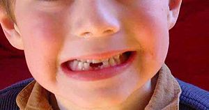
A young boy after losing two baby teeth, exfoliated in response to the permanent teeth beneath, which will erupt through the gums to take their place.

Tooth loss is when one or more teeth come loose and fall out. Tooth loss is normal for deciduous teeth (baby teeth), when they are replaced by a person's adult teeth. Otherwise, losing teeth is undesirable and is the result of injury or disease, such as mouth trauma, tooth injury, tooth decay, and gum disease. The condition of being toothless or missing one or more teeth, is called edentulism.


Prevention of tooth loss

Tooth loss due to tooth decay and gum disease may be prevented by practicing good oral hygiene, and regular check-ups (twice per year) at the dentist's office.

In contact sports, risk of mouth trauma and tooth injury is reduced by wearing mouthguards and helmets with a facemask (e.g., football helmet, and goalie mask).


Missing tooth replacement

There are three basic ways to replace a missing tooth or teeth, including a fixed dental bridge, dentures, and dental implants.


Research in tooth regeneration

Researchers in Japan have successfully regrown fully functional teeth in mice. Epithelial and mesenchymal cells were extracted from the mice, cultured to produce a tooth "germ," and the germ was then implanted into the bone at the space of a missing tooth. A tooth of the correct external and internal structure, hardness, strength and sensitivity later erupted in the space, eventually meeting the opposing tooth in a manner similar to an original natural tooth. This technique may be a possible future treatment for replacement of missing teeth.


Tooth abscess

A tooth abscess or root abscess is pus enclosed in the tissues of the jaw bone at the tip of an infected tooth. Usually the abscess originates from a bacterial infection that has accumulated in the soft pulp of the tooth.

Abscesses typically originate from dead pulp tissue, usually caused by untreated tooth decay, cracked teeth or extensive periodontal disease. A failed root canal treatment may also create a similar abscess.

There are three types of dental abscess. A gingival abscess that involves only the gum tissue, without affecting either the tooth or the periodontal ligament. A periapical abscess starts in the dental pulp. A periodontal abscess begins in the supporting bone and tissue structures of the teeth.


Presentation and symptoms

"The main symptom is a severe toothache. The pain is continuous and may be described as gnawing, sharp, shooting, or throbbing." Putting pressure or warmth on the tooth may induce extreme pain. There may be a swelling present at either the base of the tooth, the gum, and/or the cheek.

A chronic abscess may be painless but still have a swelling present on the gum. It is important to get anything that presents like this checked by a dental professional as it may become acute later.

In some cases, a tooth abscess may perforate bone and start draining into the surrounding tissues creating local facial swelling. In some cases, the lymph glands in the neck will become swollen and tender in response to the infection. It may even feel like a migraine as the pain can transfer from the infected area. The pain does not normally transfer across the face, only upwards or downwards as the nerves that serve each side of the face are separate.


Treatment

In the short term, the topical application of oil of cloves, which contains Eugenol, to the infected area is well-documented as an effective remedy.

Successful treatment of a dental abscess centers on the reduction and elimination of the offending organisms. If the tooth can be restored, root canal therapy can be performed. Nonrestorable teeth must be extracted, followed by curettage of all apical soft tissue.

Unless they are symptomatic, teeth treated with root canal therapy should be evaluated at 1- and 2-years intervals to rule out possible lesional enlargement and to ensure appropriate healing.

Abscesses may fail to heal for several reasons:

  • Cyst formation
  • Inadequate root canal therapy
  • Vertical root fractures
  • Foreign material in the lesion
  • Associated periodontal disease
  • Penetration of the maxillary sinus

Following conventional, adequate root canal therapy, abscesses that do not heal or enlarge are often treated with surgery and filling the root tips; and will require a biopsy to evaluate the diagnosis.


Untreated consequences

If left untreated, a severe tooth abscess may become large enough to perforate bone and extend into the soft tissue eventually becoming osteomyelitis and cellulitis respectively. From there it follows the path of least resistance and may spread either internally or externally. The path of the infection is influenced by such things as the location of the infected tooth and the thickness of the bone, muscle and fascia attachments.

External drainage may begin as a boil which bursts allowing pus drainage from the abscess, intraorally (usually through the gum) or extra orally. Chronic drainage will allow an epithelial lining to form in this communication to form a pus draining canal (fistula). Sometimes this type of drainage will immediately relieve some of the painful symptoms associated with the pressure.

Internal drainage is of more concern as growing infection makes space within the tissues surrounding the infection. Severe complications requiring immediate hospitalisation include Ludwig's angina, which is a combination of growing infection and cellulitis which closes the airway space causing suffocation in extreme cases. Also infection can spread down the tissue spaces to the mediastinum which has significant consequences on the vital organs such as the heart. Another complication, usually from upper teeth, is a risk of septicaemia (infection of the blood), from connecting into blood vessels, brain abscess, (extremely rare) or meningitis, (also rare).

Depending on the severity of the infection, the sufferer may feel only mildly ill, or may in extreme cases require hospital care.


Bruxism


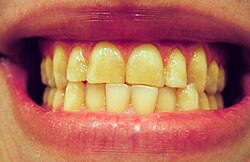
A profile of a smile, exhibiting significant wear, especially on the maxillary incisors. Even though the teeth are in an edge-to-edge position, the teeth are in maximum intercuspation; this patient possesses a Class III occlusion.

Bruxism (from the Greek βρυγμός (brugmós), "gnashing of teeth") is characterized by the grinding of the teeth and is typically accompanied by the clenching of the jaw. It is an oral parafunctional activity that occurs in most humans at some time in their lives. In most people, bruxism is mild enough not to be a health problem. While bruxism may be a diurnal or nocturnal activity, it is bruxism during sleep that causes the majority of health issues and can even occur during short naps. Bruxism is one of the most common sleep disorders.

Etiology

Numerous articles have incorrectly cited bruxism as being a reflex chewing activity, but bruxism is more accurately classified as a habit. Reflex activities happen reliably in response to a stimulus, without involvement of subconscious brain activity, and bruxism does not. All habitual activities are triggered by one kind of stimulus or another, and that does not make the habit a reflex. Chewing is a complex neuromuscular activity that is controlled by subconscious processes, with higher control by the brain. During sleep, the subconscious processes become active, while the higher control is inactive, resulting in bruxism. Some bruxism activity is rhythmic (like chewing), and some is sustained (clenching). Researchers classify bruxism as "a habitual behavior, and a sleep disorder."

The etiology of problematic bruxism is unknown, though several conditions are known to be linked to bruxism. It is theorized that certain medical conditions can trigger bruxism, including digestive ailments and anxiety.

Signs

The effects of bruxism on an anterior tooth, revealing the dentin and pulp which are normally hidden by enamel

Most bruxers are not aware of their bruxism, and only 5% go on to develop symptoms, such as jaw pain and headaches, which will require treatment. In many cases, a sleeping partner or parent will notice the bruxism before the person experiencing the problem becomes aware of it.

Bruxism can result in abnormal wear patterns of the occlusal surface, abfractions and fractures in the teeth. This type of damage is categorised as a sign of occlusal trauma.

Over time, dental damage will usually occur. Bruxism is the leading cause of occlusal trauma and a significant cause of tooth loss and gum recession.

In a typical case, the canines and incisors of the opposing arches are moved against each other laterally, i.e., with a side-to-side action, by the medial pterygoid muscles that lie medial to the temporomandibular joints bilaterally. This movement abrades tooth structure and can lead to the wearing down of the incisal edges of the teeth. People with bruxism may also grind their posterior teeth, which will wear down the cusps of the occlusal surface. Bruxism can be loud enough to wake a sleeping partner. Some individuals will clench the jaw without significant lateral movements. Teeth hollowed by previous decay (caries), or dental drilling, may collapse, as the cyclic pressure exerted by bruxism is extremely taxing on the tooth structure.


Symptoms

Patients may present with a variety of symptoms, including:

  • Morning migraines
  • Morning headaches
  • Jaw pain
  • TMJ pain
  • Facial muscle and nerve pain
  • Earache
  • Sinus pain
  • Tinnitus (ringing in the ear)
  • Vertigo
  • Neck pain
  • Shoulder pain
  • Back pain
  • Poor sleep
  • Waking exhausted
  • Stress or tension
  • Depression
  • Eating disorders
  • Insomnia
  • Daytime sleepiness
  • Eye irritation
  • Head tingling

Sequelae

Eventually, bruxism shortens and blunts the teeth being ground and may lead to myofacial muscle pain, temporomandibular joint dysfunction and headaches. In severe, chronic cases, it can lead to arthritis of the temporomandibular joints. The jaw clenching that often accompanies bruxism can be an unconscious neuromuscular daytime activity, which should be treated as well, usually through physical therapy (recognition and stress response reduction).


Diagnoses

Bruxism can sometimes be difficult to diagnose by visual evidence alone, as it is not the only cause of tooth wear. Over-vigorous brushing, abrasives in toothpaste, acidic soft drinks and abrasive foods can also be contributing factors, although each causes characteristic wear patterns that a trained professional can identify. Additionally, the presenting symptoms may be difficult for a physician to attribute to bruxism.

The effects of bruxism may be quite advanced before sufferers are aware they brux. Abraded teeth are usually brought to the patient's attention during a routine dental examination. If enough enamel has been abraded, the softer dentin will be exposed, and abrasion will accelerate. This opens the possibility of dental decay and tooth fracture, and in some people, gum recession. Early intervention by a dentist is advisable.

The most reliable way to diagnose bruxism is through EMG (electromyographic) measurements. These measurements pick up electrical signals from the chewing muscles (masseter and temporalis). This is the method used in sleep labs. There are three forms of EMG measurement available to consumers for use outside sleep labs. The first is bedside EMG units similar to those used by sleep labs. These units can be purchased for about $2000 and pick up their signals from facial muscles through wires connecting the bedside unit to electrodes that are adhesively attached to the user's face. TENS electrodes or ECG electrodes may be used.

The second type of EMG measurement available to consumers is a self-contained EMG measurement headband sold under the trade name SleepGuard, available on loan from some dentists or at a rental rate of $50 per month from the manufacturer. The EMG measurement headband does not require adhesive electrodes or wires attached to the face. While it does not record the exact time, duration, and strength of each clenching incident as the most expensive bedside EMG monitors do, it does record the total number of clenching incidents and the total clenching time each night. These two numbers easily distinguish clenching from rhythmic grinding and allow dentists to quantify severity levels accurately.

Bedside EMG units and the self-contained EMG measurement headband can both be used either in silent mode as a diagnosis measurement or in biofeedback mode as a treatment.

A third method of diagnosis using EMG is available in disposable form under the trade name BiteStrip. The BiteStrip is a self-contained EMG module that adhesively mounts to the side of the face over the masseter muscle. The BiteStrip can only do one night of measurement and does not display the clench count or total clenching time, but rather provides a single-digit display related to bruxism severity. The BiteStrip provides significantly less information than an EMG bedside unit or EMG headband and costs about $60 per day to use.

Associated factors

The following factors are associated with bruxism.

  • Disturbed sleep patterns and other sleep disorders (obstructive sleep apnea, hypopnea, snoring, moderate daytime sleepiness)
  • Malocclusion, in which the upper and lower teeth occlude in a disharmonic way, e.g., through premature contact of back teeth[citation needed]
  • Relatively high levels of consumption of caffeinated drinks and foods, such as coffee, colas, and chocolate
  • High levels of blood alcohol
  • Smoking
  • High levels of anxiety, stress, work-related stress, irregular work shifts, stressful profession and ineffective coping strategies
  • Drug use, such as SSRIs and stimulants, including methylenedioxymethamphetamine (ecstasy), methylenedioxyamphetamine (MDA), methylphenidate and other amphetamines, including those taken for medical reasons
  • Hypersensitivity of the dopamine receptors in the brain
  • GHB and similar GABA-inducing analogues such as Phenibut, when taken with high frequency
  • Disorders such as Huntington's and Parkinson's diseases
  • Obsessive Compulsive Disorder
  • Chemical drugs

Treatment

There is no single accepted cure for bruxism. However, treatments are available.

Bruxism may be reduced or even eliminated when the associated factors, e.g., sleep disorders, are treated successfully.

Mouthguards and splints

Ongoing management of bruxism is based on minimizing the abrasion of tooth surfaces by the wearing of an acrylic dental guard, or splint, designed to the shape of an individual's upper or lower teeth from a bite mould. Mouthguards are obtained through visits to a dentist for measuring, fitting, and ongoing supervision. There are four possible goals of this treatment: constraint of the bruxing pattern such that serious damage to the temporomandibular joints is prevented, stabilization of the occlusion by minimizing the gradual changes to the positions of the teeth that typically occur with bruxism, prevention of tooth damage, and the enabling of a bruxism practitioner to judge—in broad terms—the extent and patterns of bruxism through examination of the physical indentations on the surface of the splint. A dental guard is typically worn on a long-term basis during every night's sleep. Although mouthguards are a first response to bruxism, they do not in fact help cure it. An otc soft mouthguard is not considered effective by most. These mouthguards can cost anywhere from $200 to $400.

Professional treatment is medically recommended to ensure proper fit, make ongoing adjustments as needed, and check that the occlusion (bite) has remained stable. Monitoring of the mouthguard is suggested at each dental visit.

Another type of device sometimes given to a bruxer is a repositioning splint. A repositioning splint may look similar to a traditional night guard, but is designed to change the occlusion, or bite, of the patient. Randomly controlled trials with these type devices generally show no benefit over more conservative therapies, and they should be avoided under most, if not all, circumstances.

The NTI-tss device is another option that can be considered. The NTI covers only the front teeth and prevents the rear molars from coming into contact, thus limiting the contraction of the temporalis muscle. The NTI must be fitted by one's dentist.

The efficacy of such devices is debated. Some writers propose that irreversible complications can result from the long-term use of mouthguards and repositioning splints.

Biofeedback

Various biofeedback devices are currently available, and effectiveness varies significantly depending on whether the biofeedback is used only during waking hours, or during sleep as well. Many authorities remain unconvinced of the efficacy of daytime-only biofeedback. The efficacy of biofeedback delivered during sleep can depend strongly on daytime training, which is used to establish a Pavlovian response to the biofeedback signal that persists during sleep.

The first wearable nighttime bruxism biofeedback device (a biofeedback headband introduced in 2001) was originally sold under the trademark GrindAlert by BruxCare and is now sold under the GrindAlert and SleepGuard trademarks by Holistic Technologies, which holds an exclusive worldwide license to the technology. The biofeedback headband is a battery-powered device that sounds a tone against the forehead when it senses EMG (electromyographic) muscle activity in the temporalis muscles. This device records and displays nightly data on the number of bruxism events that last for at least two seconds and the total accumulated duration of those events. The volume of the alarm and the bite force required to trigger the device are adjustable. After proper Pavlovian training during waking hours, more than 25% of users achieve significantly reduced bruxism. The biofeedback sound on the headband is designed to come on slowly, allowing users to subconsciously respond in their sleep without waking up. The manufacturer offers a free three-week trial so that only people who find the device works well for them have to pay for it and claims that less than 15% of trial units are returned.

A mild electric shock bio-feedback device for treating Bruxism, GrindCare, has been approved by the European regulatory authorities and was introduced to the market in 2008. The GrindCare device infringes the US patents on the SleepGuard/GrindAlert device, so the GrindCare device is not legal to import into the United States without a license from Holistic Technologies. The device works by using simple electrodes mounted on the skin close to the cheek bones prior to sleeping; it detects the initial muscular contractions and immediately provides insignificant pulses to the facial muscles, whereby the contractions are stopped. The device is worn on the head and reportedly reduces grinding without interfering with the sleep of the patient as described by Jadidi, Castrillon & Svensson. Thereby, facial tension, joint defects and teeth disruption are reportedly reduced.

A taste-based biofeedback method was developed by Moti Nissani, Ph.D. and is called "The Taste-Based Approach to the Prevention of Teeth Clenching and Grinding". The therapy involves suspending sealed packets containing a bad-tasting substance (e.g. hot sauce, vinegar, denatonium benzoate, etc.) between the rear molars using an orthodontic-style appliance. Any attempt to bring the teeth together will rupture the packets and alert the user to the habit. This approach finds favor with some people who prefer to relate to biofeedback as "aversive therapy". The Taste-Based Approach claims to suffer less from desensitization over time than sound-based biofeedback approaches may have, but may interrupt sleep more. (There is effectively no limit to the aversive taste of certain substances. We can therefore be sure that some harmless substance exists that will alert anyone to the habit.)

One bruxism biofeedback device which was briefly on the market but is no longer available was sold under the trademark Oralsensor. This device consisted of a pneumatic pouch embedded in a soft polymer plate that fits over upper or lower teeth. When the teeth came together with a force that exceeded a set threshold, an alarm is sounded in an earpiece worn by the user; the device is no longer sold.

In 2005, a new type of occlusive device was patented that produces a movement incompatible with teeth clenching. When nighttime bruxism occurs, people breathe through the nose. The device forces people to breathe through the mouth; by forcing the opening of the mouth, the device is claimed to stop clenching. The occlusive device has an electromyogram system that monitors the electric activity of the jaw muscle via wireless electrodes. These electrodes transfer jaw-muscle activity by radio frequency to an external monitoring system. Once the signal has been interpreted by the monitoring system, if a person clenches, the monitoring unit sends a radio frequency signal to a transceiver integrated in a mechanical actuator. The mechanical actuator has two occlusive flaps that block the nostrils, forcing breathing to occur through the mouth. Once the patient stops clenching, the flaps open, allowing breathing through the nose again. The occlusive device does not wake up people since it blocks nostrils slowly, and it never closes them completely to avoid sleep disruption.

Botox

Botulinum toxin (Botox) can be successful in lessening effects of bruxism, though serious side-effects are possible. Less than one microgram ingested or inhaled is sufficient to kill an adult human. In extremely dilute form (Botox), this toxin is used as an injectable medication that weakens (partially paralyzes) muscles and has been used extensively in cosmetic procedures to relax the muscles of the face and decrease the appearance of wrinkles. In April, 2008, a study was published in the Journal of Neuroscience that showed that facially injected Botox can and does propagate into the brains of some test animals, and the U.S. Food and Drug Administration (FDA) announced that it was beginning a safety review of Botox and other similar drugs.

Botox was not originally developed for cosmetic use. It was, and continues to be, used to treat diseases of muscle spasticity such as blepharospasm (eyelid spasm), strabismus (crossed eyes) and torticollis (wry neck). Bruxism can also be regarded as a disorder of repetitive, unconscious contraction of the masseter muscle (the large muscle that moves the jaw). In the treatment of bruxism, Botox works to weaken the muscle enough to reduce the effects of grinding and clenching, but not so much as to prevent proper use of the muscle. The strength of Botox is that the medication goes into the muscle and is not supposed to get absorbed into the body (though the new research shows it does). The procedure involves about five or six simple, relatively painless injections into the masseter muscle. It takes a few minutes per side, and the patient starts feeling the effects the next day. Occasionally, some bruising can occur, but this is quite rare. Injections must be repeated more than once per year, and the risk factor of spread of the botulinum toxin is compounded by this repetition.

The symptoms that can be helped by this procedure include:

  • Grinding and clenching
  • Morning jaw soreness
  • TMJ pain
  • Muscle tension throughout the day
  • Migraines triggered by clenching
  • Neck pain and stiffness triggered by clenching

The optimal dose of Botox must be determined for each person as some people have stronger muscles that need more Botox. This is done over a few touch-up visits with the physician injector. This treatment is expensive, but sometimes Botox treatment of bruxism can be billed to medical insurance. The effects last for about three months. The muscles do atrophy, however, so after a few rounds of treatment, it is usually possible either to decrease the dose or increase the interval between treatments.

Other authorities caution that Botox should only be used for temporary relief for severe cases and should be followed by diagnosis and treatment to prevent future bruxism or jaw clenching, suggesting that prolonged use of Botox can lead to permanent damage to the jaw muscle.

Dietary supplements

There is anecdotal evidence that suggests taking certain combinations of dietary supplements may alleviate bruxism; pantothenic acid, magnesium, and calcium are mentioned on dietary supplement websites. Calcium is known to be a treatment for gastric problems, and gastric problems such as acid reflux are known to increase bruxism.

Repairing damage

Damaged teeth can be repaired by replacing the worn natural crown of the tooth with prosthetic crowns. Materials used to make crowns vary; some are less prone to breaking than others and can last longer. Porcelain fused to metal crowns may be used in the anterior (front) of the mouth; in the posterior, full gold crowns are preferred. All-porcelain crowns are now becoming more and more common and work well for both anterior and posterior restorations. To protect the new crowns and dental implants, an occlusal guard should be fabricated to wear during sleep.